Kangle Zheng, Prasanta Kumar Subudhi, Jessica Domingo,
Gerard Maopantay and Ning Huang
Plant Breeding, Genetics and Biochemistry Division,
IRRI, P. 0. Box 933, Manila, Philippines
During the PCR-based marker assisted pyramiding of
disease resistance genes in rice, we developed a DNA isolation protocol
suitable for PCR analysis and replaced ethidium bromide by methylene blue
to visualise the DNA bands in the gel. The protocol
we used for DNA isolation does not require liquid nitrogen, needs only
very small amount of tissue sample and is very rapid. Quality of isolated
DNA from this protocol is good for PCR analysis. Satisfactory results have
been obtained from these DNAs in PCR-based analysis compared to the DNAs
extracted by the conventional large scale procedure using liquid nitrogen.
These results are reproducible and this protocol is now standard procedure
for PCR-based DNA marker-assisted breeding at IRRI.
Plant tissues at different growth stages from young
seedling to maturity can be used for DNA isolation. A healthy leaf blade
(about 2 cm long) is collected in 1.5 ml tube. The tube is capped and placed
on ice. The leaf tissue is cut into half cm long and placed in a well of
spot test plate (Thomas Scientific). Four hundred microliters of extraction
buffer (Tris-HCL 50 mM, pH 8.0, EDTA 25 mM, NaCl 300 mM, SDS 1 %) is added.
The tissue is ground using a thick glass rod as a pestle. Again 400 microliters
of the extraction buffer added, mixed and 400microliters of it is transferred
into the original 1.5ml tube. Then 400microliter chloroform (containing
4% V/V isoamyl alcohol) is added and mixed well, spun for 30 seconds in
microcentrifuge. Care is taken not to disturb the interface. The supernatant
is transferred to another 1.5ml tube. To the supernatant, 800 microliter
absolute alcohol is added and mixed gently. The tube is spun for 3 min
in microcentrifuge with full speed and the supernatant is decanted. The
pellet is washed with 70% ethanol and air dried. DNA is suspended in 50microliter
of TE (Tris-HCI 10 mM, pH 8.0, EDTA 1 mM) and then stored at -20°C.
The DNA can be directly used in PCR without quantification. One microliter
of DNA is used for a PCR analysis. PCR conditioins for STSs (Sequence tagged
sites) was similar to those reported by Hittalmani et al. (1995).
One microliter of DNA was added to 24microliter of PCR mix (10 mM Tris-HCI,
pH 8.4, 50 mM KCI, 1.8 mM MgCl2, 0.01% gelatin, 100 microM dNTPs, 50 ng
of each of the primers and 1 unit of Taq DNA polymerase). Template DNA
was initially denatured at 94°C for 2 min followed by 30 cycles of
94°C for 30 sec, 55°C for 30 sec and 72°C for 1 min with a
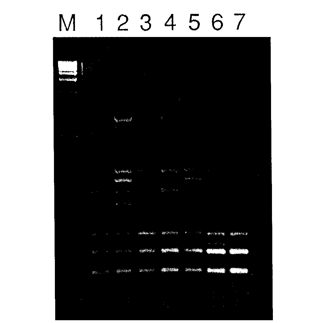
final extension step at 72°C for 5 min. Fig. 1 shows the PCR product
amplified from DNAs isolated using the above protocol from different tissues
at different growth stages with a pair of primers of a STS linked to bacterial
leaf blight resistance gene Xa-21 (Chunwongse et al. 1993).
RAPD (Random Amplified Polymorphic DNA) analysis
was performed following the protocol of Williams et a/.(1990) with
minor modification. Amplification reactions were carried out in 25 microliters
containing 10 mM Tris-HCI pH 8.3, 50 mM KCI, 0.01% (W/V) gelatin, 1.9 mM
MgCl2, 100microM each of dATP, dCTP, dGTP and dTTP, 40 ng of primer, 1
microliter of rapidly isolated DNA and 1 units of Taq polymerase.
Amplification profile was 94°C for 2 minutes, followed by 45 cycles
of 1 min at 94°C, 1 min at 36°C, 2 min at 72°C. with a final
extension of 7 min at 72°C. Amplification products were separated in
1.5% agarose gel in IX TBE buffer at 5V/cm for 5 hours. Fig. 2 shows the
RAPD amplification products of 5 BC1 progenies along with the
parents from the cross BG 309/BS1206/BS1206. Currently we are using this
miniscale protocol for RAPD screening of backcross derived plants in our
marker-assisted backcross breeding program.
Staining DNA in agarose gels with methylene blue
is basically according to Micklos and Freyer (1990). The major advantage
of using methylene blue as alternative to

Fig. 1. PCR analysis of DNA rapidly isolated from 5 different rice tissues; young leaf (1), old green leaf (2), green panicle (3), panicle before flowering (4) and root (5). The analysis was repeated twice with PCR primers derived from Chunwongse et al. (1993). Molecular weight marker (M) is Kb ladder from BRL. The gel was stained with ethidium bromide.
References
Chunwongse, J., G. B. Martin and S. D. Tanksley, 1993. Pregermination
genotypic screening using RCR
amplification of half-seeds.
Theor. Appl. Genet. 86: 694-698.
Hittalmani, S., M. R. Foolad, T. Mew, R. L. Rodriguez and N. Huang,
1995. Development of a PCR-based
marker to identify rice
blast resistance gene, Pi-2(t) in a segregating population. Theor.
Appl. Genet. 91: 9-14.
Micklos, D. A. and G. A. Freyer, 1990. DNA Science; A First Course
in Recombinant DNA Technology.
Carolina Biological Supply Company and Cold Spring
Harbour Laboratory Press, North Carolina, USA, pp.266.
Williams, S. G. K� A. R. Kubelik, K. J. Livak, J. A. Rafalski
and S. V. Tingey, 1990. DNA polymorphisms
amplified by arbitrary primers
are useful as genetic markers. Nucleic Acids Res. 18: 6531 -6535.
")
")
")
")
")
")
")
")

")


